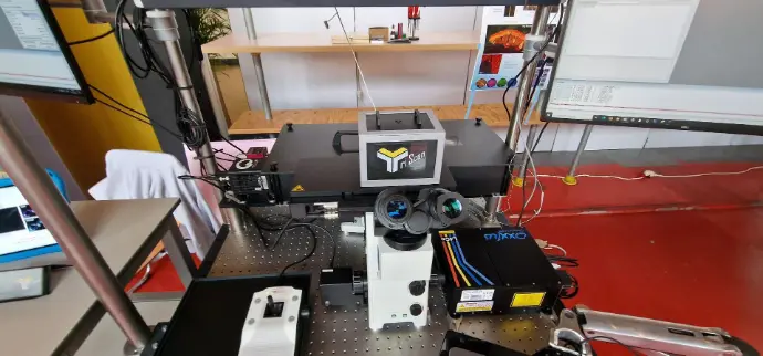
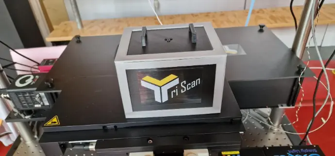
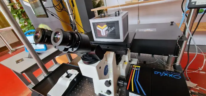
Tri scan Spinning disc System
What is the : TriScan : Tri scan Spinning disc System ?
Offering a balance of speed, sensitivity, and flexibility, the TriScan Spinning Disc System is the perfect solution for translational medicine, cancer research, and developmental biology, where confocal microscopy performance is critical.
Who it’s for
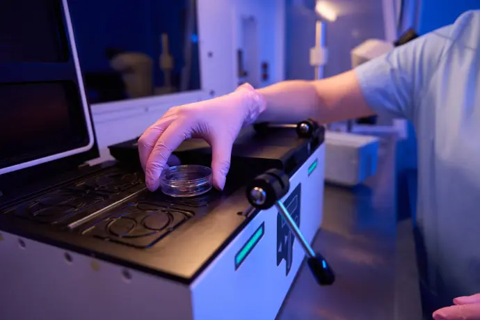
The system is designed for life sciences laboratories, core imaging facilities, academic research institutes, and biotechnology environments, providing powerful tools for researchers in cell biology, neuroscience, immunology, microbiology, and developmental biology.

✽ What We Offer
Key Technical Benefits
Ultra-Fast Confocal Imaging
TriScan employs a multi-beam scanning principle, allowing simultaneous illumination and detection of multiple points across the sample.
This results in frame rates significantly higher than traditional single-beam confocal systems, while preserving optical sectioning quality.
Superior Light Efficiency
Innovative optical design ensures higher photon collection efficiency, crucial for imaging dim fluorophores.
Reduced excitation exposure translates into lower phototoxicity for sensitive live samples.
Flexible Laser Integration
Compatible with multiple laser lines (visible and near-infrared).
Enables multi-color imaging for complex biological experiments.
Optimized for Live-Cell Studies
High-speed scanning reduces sample stress.
Efficient cooling and stable optics maintain long-term imaging performance.
Applications
Neuroscience: High-speed calcium imaging, synaptic activity mapping, and 3D reconstructions of neuronal networks.
Cell Biology: Dynamic processes such as vesicle trafficking, cytoskeletal remodeling, and cell division.
Developmental Biology: Imaging embryonic development with minimal photodamage.
Biomedical Research: Tumor microenvironment analysis, immunology, and organoid imaging.
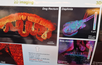
Images have been binned 4×
TriScan vs. Spinning disc Confocal Microscopes
| Feature | TriScan | Nikon A1R HD / AX |
|---|---|---|
| Scanning Mode | Multi-beam parallel scanning | Single-beam resonant or galvano scanning |
| Speed | Extremely high (ideal for fast dynamics) | High (resonant scanning), but slower for large FOV |
| Phototoxicity | Very low due to reduced exposure | Higher with long acquisitions |
| Spectral Flexibility | High (supports multiple fluorophores) | High |
| Ease of Live-Cell Imaging | Optimized for continuous fast imaging | Good, but risk of bleaching in long recordings |
Both TriScan and Nikon systems offer high-quality confocal imaging, but TriScan stands out when speed and live-cell compatibility are critical.
Why Choose TriScan?
Unmatched Imaging Speed – Capture rapid biological events with minimal delay.
Reduced Photodamage – Preserve cell viability for long-term experiments.
Flexible Integration – Works with standard inverted microscopes and multiple laser sources.
Cost-Effective Alternative – Provides confocal performance at lower maintenance and operating costs compared to large commercial systems.
Support & Training
Installation & Alignment
Professional installation by certified specialists
Full alignment and calibration for optimal performance
Step-by-step setup guidance for laboratory integration

User Training
On-site training sessions for research teams
Online tutorials and remote workshops
Covers imaging workflows: z-stack, 3D fluorescence, quantitative analysis

Technical Support
Dedicated support team for troubleshooting
Rapid response to minimize downtime
Remote diagnostics and assistance available

Software Updates & Upgrades
Regular updates for stability and performance
Access to the latest features and image analysis tools
Compatibility with new probes, biosensors, and applications

Long-Term Value
Continuous training opportunities
Extended service contracts available
Reliable support for academic, core, and industrial facilities
The Tri scan Spinning disc System bridges the gap between traditional laser scanning confocals and spinning-disk technology, delivering fast, efficient, and flexible imaging for advanced life-science research. By combining high speed with low phototoxicity, TriScan enables researchers to see more, for longer, with greater confidence.
For laboratories looking to upgrade their imaging capabilities, TriScan represents a next-generation solution that is both powerful and practical.